
Acute treatment
In order to distinguish between a stroke and a brain haemorrhage, a CT scan or MRI of the head needs to be done. Normally, the CT scan is done first. This is done shortly after the patient is admitted to hospital.
On the first images, blood clots do not always show but haemorrhages are usually visible straightaway. (CT is short for computerised tomography, which means that X-rays are used to make cross-sectional images of the brain which the doctors examine. MRI means magnetic resonance imaging. These images are made using magnetic fields.)
Treatment for blood clot
If you are admitted to hospital within four and a half hours after you develop symptoms and the CT images show that there is no haemorrhage, thrombolysis treatment (treatment to break down the blood clots) should be evaluated. Thrombolysis is administered intravenously and can help reduce the damage to the brain cells by breaking down the blood clots. In some cases, a thrombectomy may be considered. This is a surgical procedure where the blood clot is removed by means of a catheter. These treatments are not right for everyone and each case is assessed individually. Thrombectomies are best suited to removing large blood clots that are not too deep. If you cannot be given this sort of acute treatment, you will be given water-soluble acetylsalicylic acid (in Norway, Dispril) to prevent new blood clots from forming.
Even if the blood clot has reduced the blood supply to parts of the brain tissue, the brain tissue can be saved if the clot is removed and you receive optimum treatment in the acute phase. This is why it is essential that you are treated quickly. Big blood clots can sometimes lead to the build-up of high pressure inside the head because of the swelling around the injured area. In some cases, it is decided to remove part of the cranium to give the swelling more space and put this back later when the swelling has gone back. This is called a hemicraniectomy.
Treatment for haemorrhage
Controlling and monitoring the blood pressure are important parts of the treatment. Sometimes patients are given drugs to manipulate the blood’s ability to coagulate, to stop the bleeding faster. On rare occasions, surgeons decide to operate to remove the blood that has accumulated and reduce the pressure inside the cranium. If the patient has a subarachnoid haemorrhage (SAH) and there is a bulge in a blood vessel (an aneurysm), this is often treated surgically. The surgeons insert a little metal clip above the bulge, or introduce a catheter into the bulge, which they fill with metal wires (this is known as aneurysm coiling).
Stroke units
Treatment in a stroke unit is recommended for everybody who has a stroke. Stroke units have interdisciplinary teams with nurses, health workers, doctors, physiotherapists, occupational therapists, and speech therapists with expertise in stroke treatment. The stroke unit must be at a fixed location in a medical, geriatric, or neurological ward. Treatment at stroke units is a combination of acute medical treatment, monitoring patient progress, complication prevention, assessment, and rehabilitation.
The following parameters are monitored closely: blood pressure, oxygen levels, and blood sugar. If the patient’s condition changes, the monitoring is adjusted. The heart rhythm needs to be monitored for at least 24 hours. Any deterioration or new strokes must be identified and treatment steps taken. If there are complications such as difficulties swallowing and impaired bladder function, they need to be assessed and appropriate actions taken. Preventing complications can be decisive for the stroke outcome.
Assessment
The most important thing is to find out why the bleeding or infarction occurred.
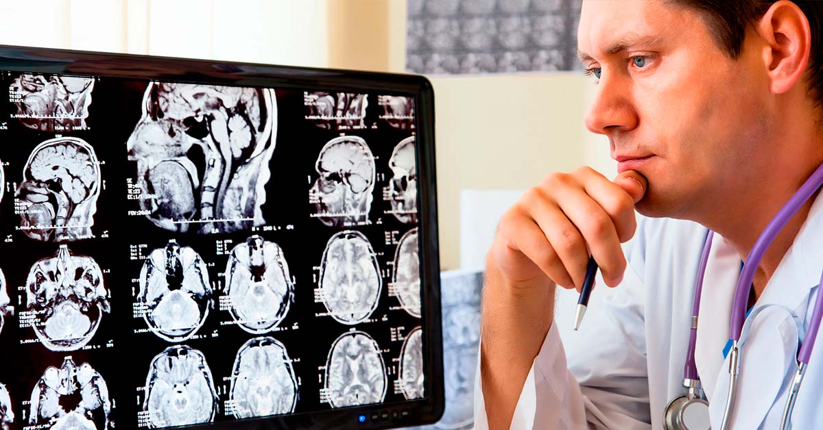
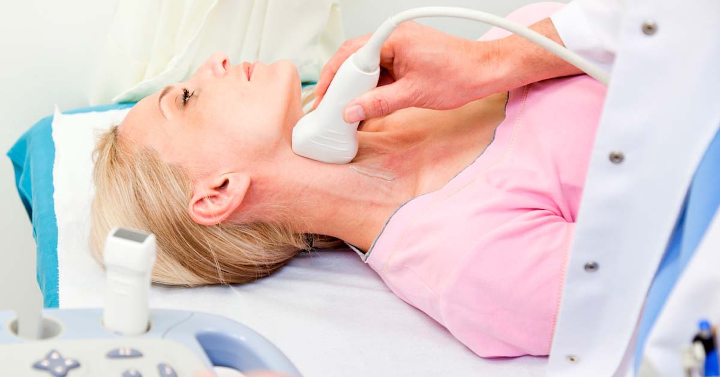
CT and/or MRI imaging of the head can show bulges in the blood vessel walls, or other factors that can have caused the brain haemorrhage. If there is a blood clot, the medical team may look into where the clot came from. Ultrasound is used to examine the carotid arteries to identify any plaque or hardening of the vessel walls that could affect the blood flow to the brain. The heart rhythm is monitored over time and the heart may be examined with ultrasound. It is possible that atrial fibrillation, changes in the septum of the heart, or any other factors in the heart that could increase the risk of a stroke will be examined. Blood tests will also be taken.
Based on the assessment, the medical team will be able to give you the right treatment or preventive medication.